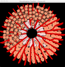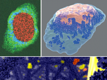
Segmentation of 3D cell tomography at cellular resolution
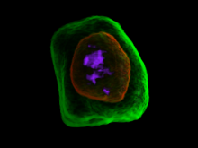
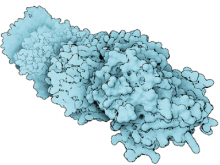
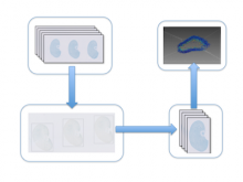
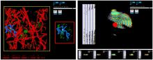
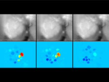
An efficient numerical algorithm is proposed and demonstrated for the segmentation of a single cell tomography with an 1- micrometer isotropic resolution. The boundaries of cell membrane and nucleus can be identified semi-automatically. The 3D image data was acquired from an ultrahigh resolution full-field optical coherence tomography (OCT) system at a rate of 160 frame/sec. The signal of OCT is coming from the scattering due to refractive index change of a skin cell (melanocyte). The OCT signal intensity is proportion to the amount of refractive index difference around the scanning points. Actual scanning data showed degradation signal-to-noise ratio at deep penetration. Using traditional methods to define clear boundaries is very difficult. Gaussian filter removes high spatial frequency signal but also removes edges. Vague boundaries can be seen by adjusting the transfer function of volume rendering, but the segmentation result changes upon the transfer function, and thus been considered unreliable. Bilateral filter was adopted to remove background noise but still retained the edge signal from the nucleus and cell membrane of the melanocyte. Here Rayburst Sampling is developed to select seeds and generate Rayburst kernel randomly within the nucleus, and to locate 3D spatial locations of the boundaries of cell membrane and nucleus as point clouds. The whole volume is then labeled according to these point clouds. As a summary, we successfully segmented the boundaries of the nucleus and membrane of a skin cell with high reliability. The depth dependent signal degradation is insignificant to the algorithm.
BioVis 2013 Information